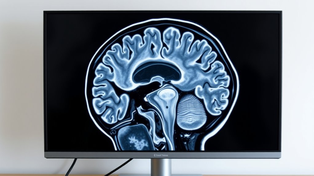
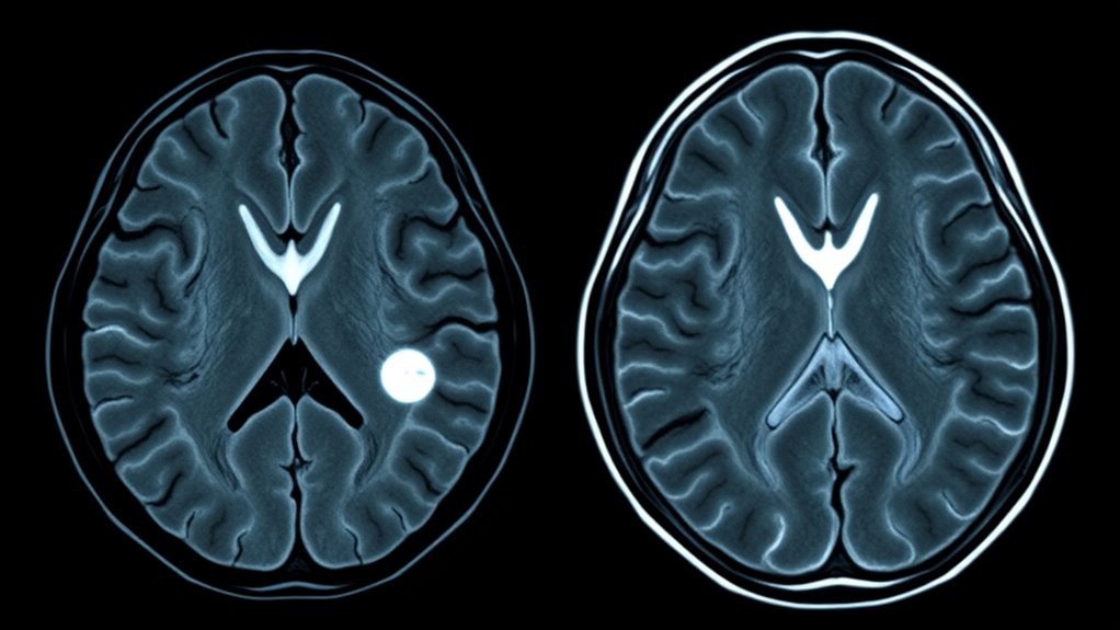
MRI helps you differentiate acoustic neuromas from other tumors by revealing their distinct features, such as location at the cerebellopontine angle and clear contrast enhancement. It shows how the tumor relates to nearby nerves and structures, highlighting specific shapes and patterns that set it apart from meningiomas or metastases. Understanding these differences allows for accurate diagnosis and treatment planning. Keep exploring to discover more about how MRI can assist in identifying these tumors precisely.
Key Takeaways
- MRI shows acoustic neuromas as well-defined, contrast-enhancing masses at the cerebellopontine angle, aiding differentiation from other tumors.
- It highlights specific location and shape features unique to acoustic neuromas versus meningiomas or metastases.
- MRI visualizes tumor relationships with nerves, vessels, and brainstem, helping distinguish origin and growth patterns.
- Different enhancement patterns and tissue characteristics on MRI help differentiate acoustic neuromas from cholesteatomas or other lesions.
- Precise imaging of tumor extent and structure allows accurate diagnosis, preventing misclassification of skull base tumors.

Magnetic Resonance Imaging (MRI) plays a essential role in diagnosing acoustic neuroma, a benign tumor that develops on the vestibulocochlear nerve. When your healthcare provider suspects this condition, MRI provides detailed tumor imaging fundamental for accurate diagnosis. The scan offers high-resolution images of the skull base, where these tumors typically grow, allowing you and your doctor to see the precise location and size of the growth. Unlike other imaging techniques, MRI excels at differentiating soft tissues, making it the preferred method for identifying acoustic neuromas.
MRI is essential for accurately diagnosing and locating acoustic neuromas at the skull base.
Because acoustic neuromas originate near critical structures like the brainstem and cerebellum, your doctor needs to distinguish them from other tumors that may develop in the same area. MRI helps do this by revealing specific characteristics of the tumor. For instance, an acoustic neuroma usually shows as a well-defined, contrast-enhancing mass at the cerebellopontine angle, which is a key site at the skull base. The tumor’s appearance on MRI scans differs from other tumors such as meningiomas or schwannomas in nearby regions, helping clinicians narrow down the diagnosis.
Tumor imaging with MRI also provides valuable information about the tumor’s relationship to adjacent structures. This is essential for planning treatment, whether surgical removal or radiotherapy. MRI can demonstrate if the tumor has grown into or compressed nearby nerves, blood vessels, or the brainstem. This detailed visualization guides surgeons in choosing the safest and most effective approach. It also helps monitor tumor growth over time, especially if a watch-and-wait approach is taken initially.
The ability of MRI to visualize the skull base is particularly important because this area houses many critical nerves and blood vessels. Clear images help to differentiate acoustic neuromas from other skull base tumors that might extend into or arise from different tissues. For example, tumors like cholesteatomas or metastases have different imaging features, and MRI helps distinguish them based on their location, shape, and enhancement patterns. This precise tumor imaging ensures you receive the proper diagnosis, avoiding unnecessary treatments or delays.
Frequently Asked Questions
Can MRI Detect Acoustic Neuroma at Its Earliest Stages?
Early detection of acoustic neuroma depends on imaging sensitivity, and MRI is highly effective for this purpose. You can benefit from MRI’s detailed views, which often reveal tumors at their smallest stages before symptoms appear. Its advanced imaging capabilities help catch these tumors early, allowing for prompt treatment. So, if you’re concerned about early detection, MRI offers a reliable, non-invasive way to identify acoustic neuromas at their earliest stages.
How Accurate Is MRI Compared to Other Imaging Techniques?
MRI is like a sharp-eyed detective, offering exceptional imaging precision and diagnostic reliability compared to other techniques. Its ability to distinguish acoustic neuroma from other tumors is highly accurate, often surpassing CT scans or ultrasound. You can trust MRI’s detailed images to provide the clearest picture, making it a crucial tool in diagnosing and planning treatment accurately. Its superior accuracy ensures you get the most reliable diagnosis possible.
Are There Risks Associated With MRI Scans for Tumor Detection?
You might wonder about MRI safety and contrast risks during tumor detection scans. Generally, MRI is safe, as it doesn’t use ionizing radiation. However, some people could experience allergic reactions to contrast agents, which carry a small risk. If you have kidney problems or allergies, tell your doctor beforehand. Overall, MRI is a safe, effective tool for identifying tumors, with minimal risks when proper precautions are taken.
How Does MRI Differentiate Benign From Malignant Tumors?
Think of MRI as your modern-day crystal ball, revealing tumor details clearly. It helps differentiate benign from malignant tumors by showing their size, shape, and location. Unlike tumor markers or biopsy methods, MRI provides a non-invasive way to evaluate tumor characteristics. It can identify features suggestive of malignancy, such as irregular borders or invasion into surrounding tissues, guiding your doctor on the best next steps for diagnosis and treatment.
What Advancements Are Being Made in MRI Technology for Tumor Diagnosis?
You’ll find that advancements in MRI technology focus on high-resolution imaging and contrast enhancement. These improvements allow you to see tumors more clearly, distinguishing their size, shape, and tissue characteristics with greater accuracy. Enhanced contrast agents help you identify tumor boundaries and differentiate benign from malignant growths more effectively. As a result, you gain more precise diagnoses, leading to better treatment planning and improved patient outcomes.
Conclusion
So, when it comes to telling acoustic neuroma apart from other tumors, MRI is your superhero. It’s like having a superpowered x-ray that reveals details no other test can match. With its precision, you can catch these tumors early and save the day. Don’t underestimate the power of MRI — it’s the ultimate detective in the world of brain tumors, making sure you get the right treatment before things get out of hand.


