
TL;DR

Get everyday helpers delivered free with Prime
- Fast, free delivery on millions of items
- Prime Video, Amazon Music and more included
- Member-only deals all year
Researchers at EPFL created a three-dimensional atlas identifying 539 chemically distinct lipid zones in mouse brains. The map also showed extensive lipid changes in pregnant mice, but it is a mouse reference, not a direct map of the human brain.
An EPFL-led research team has mapped 539 chemically distinct regions in the mouse brain by measuring how lipids are distributed across its tissues, adding a layer of detail that conventional anatomical and genetic maps can miss. The atlas, published in Nature, also records broad lipid changes in pregnant mice, giving researchers a reference for studying brain chemistry and how it varies across conditions.
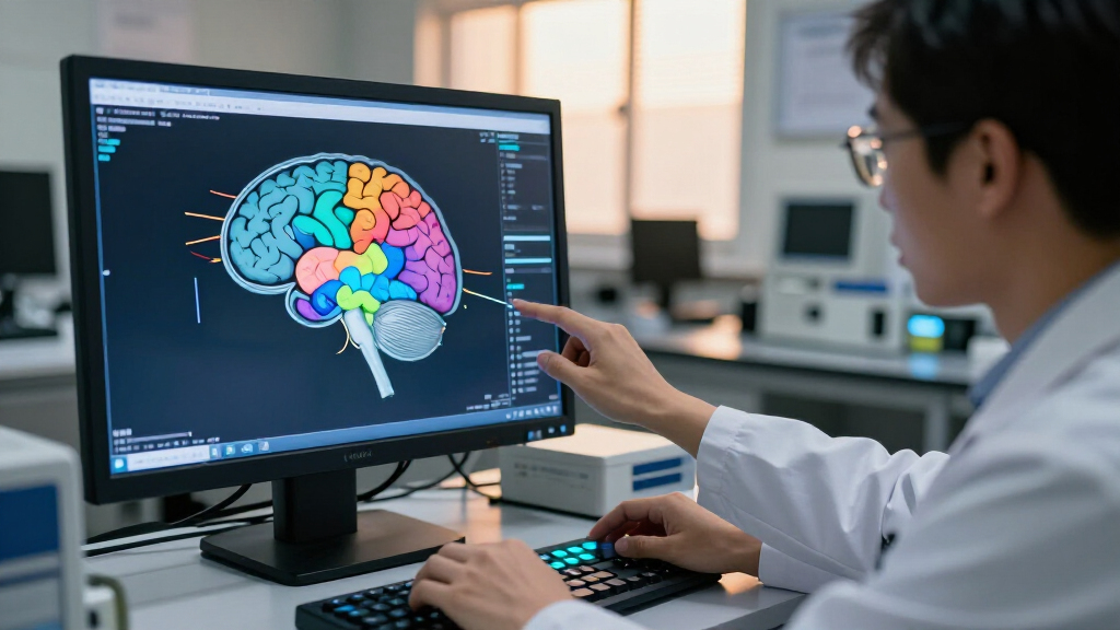
The team used mass spectrometry imaging to detect lipids in thin sections of brain tissue. Researchers analyzed 172 lipids across 109 slices from 11 mice, generating about 7 million measurements. Machine-learning tools developed for the project were used to combine those measurements into a three-dimensional reconstruction.
The resulting Lipid Brain Atlas divides the tissue into 539 “lipizones”, each defined by a characteristic combination of lipid molecules. Some zones correspond to familiar anatomical structures or cell types; others reveal chemical boundaries and relationships not readily apparent from anatomy or gene activity alone. The researchers say lipid signatures could also help identify where a tissue sample originated within the brain.
The map showed unexpected variation in white matter, which contains nerve fibers connecting brain regions. Rather than appearing chemically uniform, it contained numerous lipid-defined zones. The team also identified previously unrecognized chemical boundaries in the choroid plexus, which produces cerebrospinal fluid, and in tissues lining the brain’s internal cavities.
In a separate analysis, researchers found substantial lipid-composition changes across the brains of pregnant mice. Changes were especially widespread in the cortex, while levels of galactosyl ceramide—a lipid associated with myelin—increased sharply across much of the white matter. The report says these shifts were more pronounced overall than typical lipid differences observed between male and female mice. The study does not establish what causes the changes or what they mean for behavior or brain function.
What Lipid Zones Add to Brain Maps
Brain research often maps tissue through anatomy, cell types, connections, gene activity or proteins. The atlas adds lipid composition as another way to distinguish regions. Lipids help form cell membranes and myelin, the insulating layer around many nerve fibers, and are involved in communication between brain cells. A chemical map may therefore reveal differences in tissue that other mapping methods do not capture.
The white-matter findings illustrate that value: researchers found chemical variation in tissue that might otherwise be treated as relatively consistent. The atlas could let scientists compare lipid patterns with existing cellular and genetic maps, then investigate whether particular patterns change during development, aging or disease. Those are potential uses, not conclusions that this study has already established.
The pregnancy results offer a further research lead. They show that lipid composition in mouse brains can vary substantially with physiological state, including across regions of the cortex and white matter. The findings do not show that the same patterns occur in people, nor do they establish that the shifts directly change neural connections.
As an affiliate, we earn on qualifying purchases.
Why Brain Lipids Were Hard to Map
Scientists have extensively studied brain cells, their connections and their genes, but mapping where individual lipid molecules occur has been more difficult. According to the ScienceDaily report on the study, conventional imaging methods have struggled to distinguish individual lipids across brain tissue. That has left researchers with a less detailed picture of the brain’s chemical organization than of some other aspects of its biology.
The new project addresses that measurement problem with mass spectrometry imaging, which examines tissue one location at a time and records the molecules detected at each point. The study combines those readings across many slices rather than relying on a single image. The resulting atlas is a reference for healthy mouse brain tissue, and the researchers say the mapping approach—not the specific mouse map—could be adapted to investigate human brains.
““This shows that the cells which make myelin are far more varied in their chemistry than their genes alone would suggest.””
— Luca Fusar Bassini, study co-author
Limits of the Mouse Atlas
The atlas describes mouse brains, and the researchers caution that its map cannot be directly applied to humans. Whether people have comparable lipid-defined zones, and how those zones might differ, remains to be established. The work provides a method that could support human research, not a completed human brain atlas.
The pregnancy analysis also leaves open what drives the lipid changes, how long they last and whether they affect brain activity or function. The study reports changes in lipid composition but does not show that they cause pregnancy-related neurological effects. Nor does the atlas itself demonstrate that a particular lipid pattern causes or predicts Alzheimer’s disease, depression or other conditions. Those links would need separate research.
Using the Atlas in Further Studies
The Lipid Brain Atlas is available online for scientists to explore, according to the report. Researchers can use it as a baseline for mouse brain tissue, compare its lipid zones with genetic and cellular maps, and investigate whether those patterns shift with age, development, pregnancy or disease. The atlas is a research resource; the report does not describe a scheduled clinical application or a confirmed next study.
A key next step will be to test how well the approach works on human brain tissue and determine whether comparable chemical organization can be identified. Further studies will also be needed to clarify the pregnancy-related findings and assess how lipid changes relate to brain function. Until then, the reported regions and biological interpretations remain specific to the mouse research described in the paper.
Key Questions
What are the 539 regions in the new atlas?
They are lipizones: areas of mouse brain tissue distinguished by their characteristic combinations of lipid molecules. Some match known structures, while others mark chemical differences not previously identified in the same way.
How did the researchers create the map?
The team used mass spectrometry imaging to measure 172 lipids in 109 brain slices from 11 mice. Machine-learning tools combined about 7 million measurements into a three-dimensional atlas.
Does the atlas map the human brain?
No. The published map is based on mouse brain tissue. The researchers say the method could be used to look for comparable patterns in people, but the mouse map cannot be directly transferred to humans.
What did the study find about pregnancy?
Researchers reported substantial lipid changes across pregnant mice’s brains, including extensive shifts in the cortex and higher levels of galactosyl ceramide across much of the white matter. The study does not establish the cause or functional effects of those changes.
Source: rss
Halloween Picks
halloween
As an affiliate, we earn on qualifying purchases.