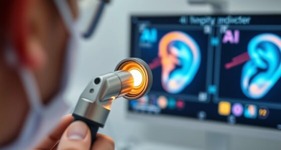
Electrocochleography (ECochG) measures the electrical responses generated by your inner ear in reaction to sound. It involves placing electrodes near your eardrum or on your tympanic membrane to record tiny signals from hair cells and nerves. These responses help diagnose issues like Ménière’s disease, cochlear dysfunction, or nerve damage. Understanding how these potentials work can provide valuable insights into your inner ear health, and if you keep exploring, you’ll find even more detailed information.
Key Takeaways
- Electrocochleography (ECochG) measures electrical potentials generated by the cochlea and auditory nerve in response to sound stimuli.
- Key signals include cochlear microphonics, summating potential, and action potentials, reflecting inner ear and neural activity.
- ECochG helps diagnose conditions like Ménière’s disease, cochlear and retrocochlear pathologies, and early inner ear dysfunction.
- Proper electrode placement, calibration, and signal interpretation are crucial for accurate decoding of inner ear potentials.
- Advances in technology, including AI analysis and wearable devices, enhance the precision and accessibility of ECochG diagnostics.
Understanding the Fundamentals of ECochG

Electrocochleography (ECochG) is a technique that records electrical responses generated by the inner ear and auditory nerve when sound stimuli are presented. To understand ECochG, you need to grasp basic inner ear physiology. The cochlea, a spiral-shaped structure, converts sound waves into electrical signals. These signals travel via the auditory nerve to your brain, allowing you to perceive sound. ECochG captures the electrical activity produced at different points along this pathway, especially the cochlear response (Cochlear Microphonics) and the activity of the auditory nerve (Summating Potential and Action Potentials). By analyzing these responses, you gain insights into how your inner ear functions and identify potential issues early. Additionally, understanding the contrast ratio of the system can aid in interpreting subtle differences in the recorded signals, enhancing diagnostic accuracy. This understanding forms the foundation for interpreting ECochG results and diagnosing auditory disorders.
The Equipment and Procedure Involved in Electrocochleography

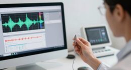
To perform ECochG, you’ll need specialized equipment that captures and amplifies the tiny electrical signals generated by the inner ear and auditory nerve. The equipment includes electrodes, an amplifier, and a data acquisition system. Electrode placement is vital; typically, a small, silver-silver chloride electrode is placed near the eardrum or on the tympanic membrane for ideal signal detection. You’ll also position a ground electrode to reduce noise. Before recording, calibration procedures verify the equipment functions correctly, adjusting for gain and impedance. Proper calibration guarantees accurate measurements. During the procedure, you’ll monitor the signals in real-time, making sure electrode contact remains stable. This setup allows precise collection of cochlear and neural responses, setting the stage for accurate interpretation of the inner ear’s electrical activity. Additionally, understanding the trustworthiness of AI models can inform the development of safer diagnostic tools.
Interpreting the Electrical Signals From the Inner Ear

Understanding how to interpret the electrical signals from the inner ear is vital for accurate diagnosis. You’ll need to recognize the mechanisms behind signal generation and apply effective clinical strategies. Mastering these points helps you distinguish normal from abnormal responses efficiently. Recognizing the importance of Inspirational Quotes About Fatherhood can be paralleled to understanding the guiding principles behind inner ear responses, providing better insight into complex audiological data.
Signal Generation Mechanisms
The electrical signals recorded during electrocochleography originate from the intricate activity of hair cells and neural pathways within the inner ear. Signal generation in the inner ear involves the transduction of sound vibrations into electrical impulses by hair cells in the cochlea. When sound waves reach the inner ear, they cause the hair cells’ stereocilia to bend, opening ion channels that generate receptor potentials. These potentials then trigger neural activity in the auditory nerve fibers. The recorded potentials, like the cochlear microphonic and summating potential, reflect these processes. Understanding how these signals are generated helps you interpret the data accurately, revealing the inner ear’s functional status. This insight is essential for diagnosing conditions like Meniere’s disease or cochlear nerve dysfunction. Additionally, the use of advanced filtration technology, such as HEPA filters, can help reduce ambient noise and improve the accuracy of signal recordings during testing.
Clinical Interpretation Strategies
Interpreting electrocochleography signals requires careful analysis of the waveform patterns and their amplitudes, as these reflect the underlying physiological processes within the inner ear. You should guarantee proper patient preparation to minimize artifacts, such as instructing the patient to relax and avoid movement. Accurate data recording techniques are essential; use consistent electrode placement and appropriate filter settings to obtain clean, reliable signals. Focus on identifying key features like the cochlear microphonic, summating potential, and action potential, comparing their amplitudes and latencies to normative data. Recognize patterns indicative of pathologies like Meniere’s disease or auditory nerve issues. Combining precise patient prep with meticulous recording ensures meaningful interpretation, enabling you to make informed clinical decisions based on the inner ear’s electrical responses. Additionally, understanding AI-driven data analysis can enhance the accuracy of waveform interpretation by identifying subtle patterns that may indicate early or hidden pathologies.
Clinical Conditions Diagnosed Using ECochG

Electrocochleography (ECochG) plays a pivotal role in diagnosing various clinical conditions related to the inner ear and auditory nerve. You can use ECochG to assist in tinnitus assessment, helping determine if abnormal inner ear activity contributes to your symptoms. It’s also essential in Ménière’s diagnosis, revealing endolymphatic hydrops that cause vertigo and hearing loss. ECochG provides objective data that supports clinical evaluations, especially when symptoms are ambiguous. Conditions you might identify include: inner ear function assessment, tinnitus and hyperacusis, Ménière’s disease, cochlear nerve disorders, and sudden sensorineural hearing loss.
Advantages and Limitations of Electrocochleography

Electrocochleography offers several key advantages that make it a useful tool in audiology and otology. It provides direct measurement of inner ear potentials, helping you identify conditions like Meniere’s disease or monitor cochlear function during surgery. The technique is relatively quick and non-invasive, making it suitable for routine assessments. However, potential discomfort can arise from electrode placement, especially if you have sensitive skin or anxiety. Additionally, cost considerations come into play, as specialized equipment and trained personnel are necessary, which may limit accessibility in some settings. Despite these limitations, the detailed insights gained from ECochG often outweigh the drawbacks, making it a valuable diagnostic tool when used appropriately. Proper self-monitoring during testing can also enhance comfort and safety for patients.
Comparing Ecochg With Other Audiological Tests

While electrocochleography (ECochG) provides direct insights into cochlear and auditory nerve function, it often complements other audiological tests rather than replacing them. It’s a valuable tool in inner ear diagnostics, especially when you need detailed information about cochlear health. Compared to other audiological testing methods, ECochG offers real-time data on inner ear potentials, making it useful for diagnosing conditions like Meniere’s disease. However, it’s usually part of a broader testing battery, including otoacoustic emissions and auditory brainstem responses. Here are some key points:
- ECochG measures electrical responses directly from the cochlea and nerve.
- It’s particularly useful for diagnosing retrocochlear and cochlear issues.
- Other audiological tests often assess hearing thresholds and outer hair cell function.
- Combining tests gives a extensive view of your inner ear health.
- Understanding inner ear potentials can enhance the interpretation of ECochG results, providing a comprehensive picture of inner ear function.
Future Developments in Inner Ear Monitoring Technologies

Advancements in technology are paving the way for more precise and less invasive inner ear monitoring methods. Future developments focus on biomarker discovery, enabling early detection of inner ear issues through blood or fluid analysis. Wearable devices are becoming more sophisticated, allowing continuous, real-time monitoring of cochlear function outside clinical settings. These innovations can improve diagnosis accuracy and patient comfort. Additionally, integrating advanced fraud detection techniques can help ensure the security of patient data collected through these new devices. Here’s a glimpse of potential tools:
| Technology | Impact |
|---|---|
| Wearable devices | Continuous monitoring of inner ear health |
| Biomarker discovery | Early detection and personalized treatment |
| AI integration | Data analysis for better diagnosis |
These advancements promise faster, more accessible inner ear assessments, transforming audiology and patient care.
Frequently Asked Questions
How Long Does an Ecochg Test Typically Take?
The procedure duration for an ecochg test usually takes about 30 to 60 minutes. During this time, you’ll sit or lie comfortably while the technician places tiny electrodes near your ear and on your forehead. The process is generally quick and non-invasive, designed to guarantee patient comfort. You might feel slight pressure from the electrodes, but overall, it’s a straightforward test that provides valuable information about your inner ear function.
Can Ecochg Detect Early-Stage Inner Ear Disorders?
You might wonder if ecochg can catch early inner ear issues. Yes, it can help identify inner ear biomarkers indicating early detection of inner ear disorders. By measuring electrical responses, ecochg detects subtle changes before symptoms appear. This makes it a valuable tool for diagnosing issues early, enabling prompt treatment. Keep in mind, though, its effectiveness depends on the specific condition and the expertise of your healthcare provider.
Is Ecochg Suitable for All Age Groups?
Thinking electrocochleography (ECOG) is suitable for everyone? Well, don’t be too quick to assume. Pediatric suitability is clear, but geriatric considerations matter—older adults might find the procedure uncomfortable or less reliable. While it’s a valuable tool, you should consider age-related factors carefully. So, yes, ECOG isn’t a one-size-fits-all solution; it’s tailored, just like your approach to each patient’s needs.
What Are the Risks or Side Effects of Ecochg?
You might experience some test discomfort during electrocochleography, such as minor irritation or pressure. Rarely, you could have allergic reactions to the electrode gels or adhesives used. While side effects are uncommon, it’s important to inform your healthcare provider if you have sensitivities. Overall, the procedure is safe, but being aware of potential discomfort or allergies helps guarantee a smooth experience.
How Does Ecochg Compare to MRI in Diagnosis Accuracy?
When comparing ecochg to MRI, you’ll find ecochg offers high diagnostic reliability for inner ear conditions, especially in detecting endolymphatic hydrops. Its comparison accuracy is quite good, but MRI provides a broader view of inner ear and brain structures, making it more extensive for certain diagnoses. Ecochg is less invasive and quicker, but MRI’s detailed images give it an edge in overall diagnostic precision for complex cases.
Conclusion
Don’t overlook electrocochleography’s potential—it’s a crucial tool for early diagnosis of inner ear issues that can be easily missed with standard tests. While some worry about its complexity, mastering ECochG offers more precise insights into your patients’ hearing health. Embracing this technology now means better outcomes and proactive care. So, why wait? Incorporate ECochG into your practice and stay ahead in audiological diagnostics.


