Modern ear exams have shifted from simple otoscope inspections to advanced digital imaging and AI analysis. You’ll notice high-resolution cameras provide clearer views of your ear canal and eardrum, while digital recordings help track changes over time. AI-powered tools analyze images instantly for quick, accurate diagnoses. This technology makes exams faster, safer, and more reliable. If you want to understand how these innovations improve your ear health evaluations, keep exploring the details behind this progress.
Key Takeaways
- Modern ear exams start with otoscopy, using specialized tools like digital otoscopes for detailed visualization of the ear canal and eardrum.
- Advanced imaging techniques, including high-resolution cameras and video otoscopy, enhance detection of abnormalities and structural issues.
- Integration of AI analyzes ear images in real-time, improving diagnostic accuracy and enabling early detection of infections or damage.
- Digital storage of images and recordings allows for better documentation, tracking, and comparison over time.
- Future innovations focus on more comprehensive, non-invasive assessments with faster, more precise, and patient-friendly examination processes.
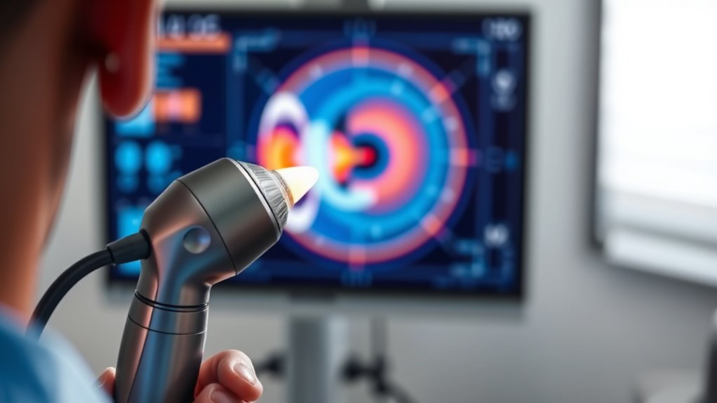
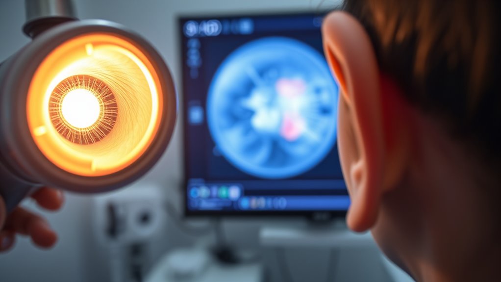
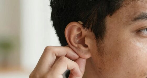
Modern ear exams have become more accurate and less invasive thanks to new technology. When you sit in front of an audiologist or ENT specialist, you might think the process involves a simple look inside your ear, but advancements have transformed how these examinations are conducted. One of the foundational steps in modern ear assessments is the ear canal examination. Using specialized tools like an otoscope, your healthcare provider can gently inspect your ear canal to check for blockages, wax buildup, infections, or signs of injury. Thanks to improved magnification and illumination, this examination is clearer and more detailed than ever before. The otoscope’s camera, often digital now, provides high-resolution images that allow for a thorough assessment without causing discomfort.
Following the ear canal examination, the tympanic membrane analysis becomes essential. The tympanic membrane, or eardrum, acts as a barrier and a transmitter of sound. With advanced imaging and visualization tools, your provider can evaluate the condition of your eardrum with confidence. They look for signs of perforation, fluid behind the membrane, or abnormal changes in its color or mobility, which can indicate infections or other issues. Modern technology allows for precise tympanic membrane analysis, often with video otoscopy, giving both the clinician and you a clear view of what’s happening inside your ear. Some devices even incorporate digital recordings, enabling detailed tracking of any abnormalities over time.
Modern imaging and video otoscopy enable precise, real-time assessment of the eardrum’s condition.
What’s impressive is how technology has streamlined these processes. Traditional ear exams relied solely on the clinician’s eye and manual tools, which could sometimes miss subtle issues. Now, digital imaging and high-definition cameras improve detection accuracy and enable better documentation. These images can be stored electronically, making follow-up assessments more consistent and reliable. Additionally, some clinics now employ AI-powered imaging systems that analyze the ear images instantly, highlighting areas of concern or abnormalities that might be overlooked by the human eye.
All of these technological innovations make ear exams faster, safer, and more precise. You don’t need to worry about discomfort or invasive procedures—most of these examinations are quick and non-invasive. They provide your healthcare provider with a thorough understanding of your ear health, helping to diagnose infections, damage, or other issues early, so appropriate treatment can begin sooner. As technology continues to advance, the process will only become more refined, ensuring you receive the best possible care during your ear health assessments.
Frequently Asked Questions
How Accurate Are Ai-Based Ear Imaging Diagnostics?
AI-based ear imaging diagnostics are highly accurate, especially when evaluating the ear canal. They improve diagnostic accuracy by providing detailed images and analysis that can detect issues missed by traditional exams. You can rely on these advanced tools to get precise results quickly, helping you identify problems early. While no technology is perfect, AI imaging considerably enhances your ability to diagnose ear conditions accurately and efficiently.
What Are the Risks of Automated Ear Examinations?
Think of automated ear exams like a GPS: reliable most of the time, but sometimes leading you astray. Risks include technology reliability issues, where errors might occur, and patient privacy concerns, if sensitive data isn’t safeguarded. You could experience misdiagnosis if the system malfunctions or if privacy isn’t maintained. Staying aware of these risks helps ensure that technology enhances your care without compromising safety or confidentiality.
Can AI Replace Traditional Otoscopy Entirely?
You might wonder if AI can fully replace traditional otoscopy. While AI offers improved sensor accuracy and can enhance diagnostic speed, it can’t yet match the nuanced judgment and patient comfort provided by a skilled examiner. AI tools are valuable supplements, but relying solely on them may overlook subtle signs. For now, combining AI with traditional methods offers the best balance of accuracy and patient care.
How Affordable Is AI Technology for Clinics?
Think of AI technology like the Wright brothers’ first flight—innovative but initially costly. For your clinic, AI imaging tools are becoming more affordable, but cost considerations still influence adoption. While prices are dropping, you must weigh benefits versus expenses carefully. Embracing this technology can streamline diagnoses and improve patient care, but guarantee your budget aligns with the potential for long-term savings and enhanced accuracy.
What Training Is Needed to Interpret AI Ear Images?
You’ll need training in machine learning basics and image annotation to interpret AI ear images effectively. This involves understanding how AI models analyze and learn from ear images, recognizing patterns, and accurately labeling features during image annotation. By gaining these skills, you can better evaluate AI-generated insights, improve diagnostic accuracy, and confidently integrate AI imaging tools into your practice.
Conclusion
Today, ear exams have transformed from simple otoscopy to advanced AI imaging, making diagnoses quicker and more accurate. You’re now equipped with tools that feel almost like magic, bringing hope and clarity where once there was uncertainty. Remember, as you embrace these innovations, you’re part of a medical revolution—like stepping into a future where technology and compassion go hand in hand, ensuring better care for every patient. The future’s bright, and you’re at the forefront of it.

