When choosing between CT and cone beam CT for temporal bone imaging, consider that traditional CT provides higher resolution images ideal for detailed microanatomy, but involves higher radiation doses and costs. Cone beam CT offers lower radiation exposure, quicker scans, and better accessibility, making it suitable for less complex cases or in pediatric patients. To guarantee you select the best option for your needs, understanding their differences can help you optimize diagnosis and treatment planning. Keep exploring to gain more insights.
Key Takeaways
- Conventional CT offers higher spatial resolution, better visualizing fine structures like tiny ossicles and inner ear components.
- Cone beam CT provides lower radiation exposure and is suitable for general assessment of temporal bone anatomy.
- Traditional CT is more effective for complex or detailed preoperative planning requiring microanatomy visualization.
- CBCT is faster, more accessible, and cost-effective, making it ideal for routine or pediatric temporal bone imaging.
- Choice depends on diagnostic needs: use CT for detailed microstructures; use CBCT for lower-dose, quicker assessments.
Overview of Imaging Modalities for Temporal Bone Analysis
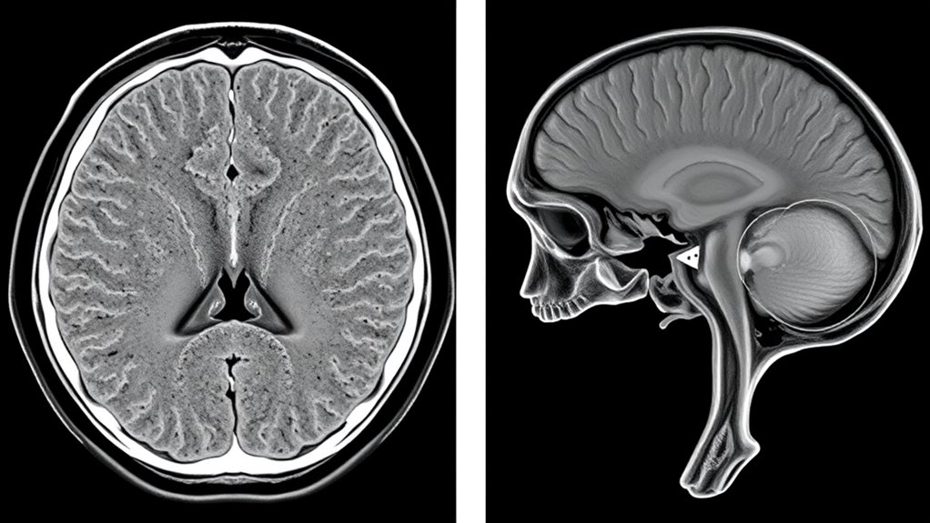
When analyzing the temporal bone, choosing the right imaging modality is crucial for accurate diagnosis and treatment planning. Both CT and cone beam CT offer detailed views, but they differ in how they handle anatomical variations. Traditional CT provides exhaustive images with high resolution, making it easier to distinguish complex structures. Cone beam CT, on the other hand, offers a more patient-friendly experience with shorter scan times and less radiation exposure. This can enhance patient comfort, especially for those who experience anxiety or have difficulty staying still. Understanding these differences helps you select the appropriate modality based on the specific anatomical variations you need to evaluate and the patient’s comfort, ensuring ideal imaging outcomes and better clinical decision-making.
Technical Aspects and Image Acquisition Processes
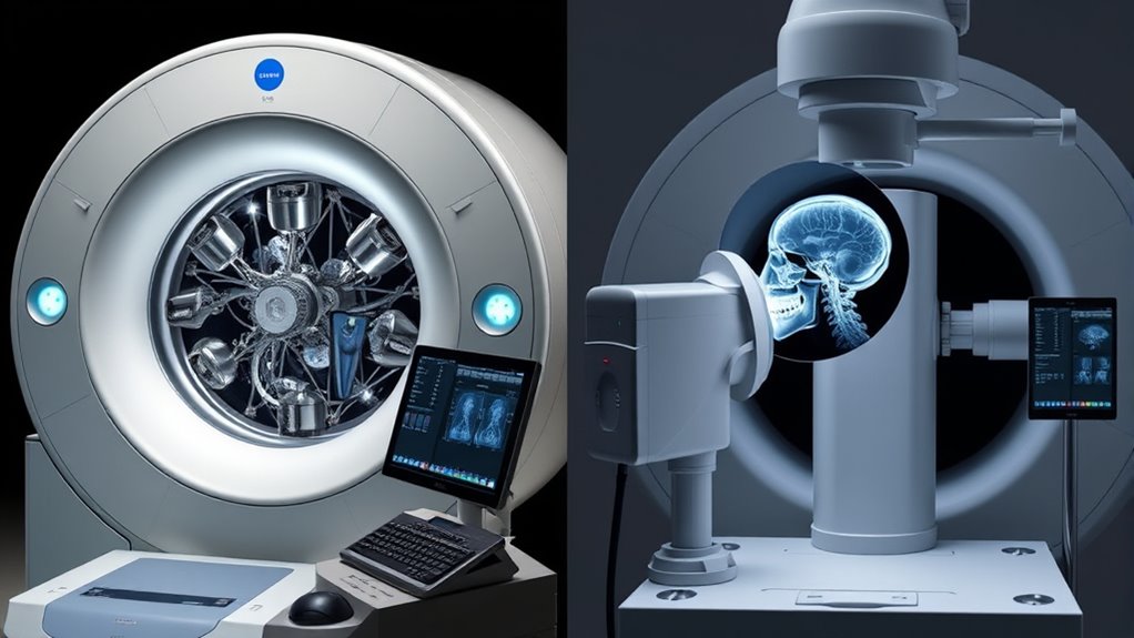
Understanding the technical aspects of image acquisition is key to enhancing results when comparing CT and cone beam CT for temporal bone imaging. Both methods rely on specific technical specifications that influence image quality and diagnostic utility. CT uses rotating X-ray beams and detectors to capture detailed cross-sectional images, with parameters like voltage (kV), current (mA), and exposure time affecting clarity and noise levels. Cone beam CT employs a cone-shaped X-ray beam and a flat-panel detector, offering rapid scans with lower radiation doses. Adjusting technical specifications such as voxel size, scan angle, and exposure settings guarantees adequate visualization of complex temporal bone structures. Mastering these technical aspects helps you attain superior image quality, enabling accurate diagnosis and effective treatment planning. Additionally, understanding the contrast ratio helps optimize image contrast and depth perception, which is crucial for detailed temporal bone evaluation.
Image Resolution and Detail Visualization
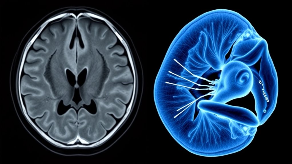
When comparing CT and cone beam CT for temporal bone imaging, understanding their spatial resolution capabilities is key. You’ll notice differences in how clearly each modality visualizes fine structures and details. This affects the overall image clarity and sharpness, impacting diagnostic confidence. Additionally, advancements in imaging technology continue to improve resolution and detail visualization in these modalities.
Spatial Resolution Capabilities
Spatial resolution plays a crucial role in imaging the complex structures of the temporal bone, directly impacting how well details are visualized. Higher spatial detail allows you to see finer structures, but resolution limits can restrict this clarity. With conventional CT, you typically get better spatial resolution, revealing small bone nuances. Conversely, Cone Beam CT offers lower resolution but covers larger areas quickly. Consider these points:
- CT provides sharper images suitable for detailed bone anatomy.
- Cone Beam CT offers sufficient spatial detail for general assessment but may miss minute features.
- Resolution limits of each modality determine how much fine detail you can trust in your diagnosis.
Understanding these differences helps you choose the right imaging technique, balancing the need for spatial detail against the limitations of resolution.
Visualizing Fine Structures
Visualizing fine structures in the temporal bone depends heavily on the image resolution of your chosen modality. Higher resolution images reveal microvascular detail, allowing you to see tiny blood vessels and delicate anatomical features clearly. This detail is essential for accurate diagnosis and surgical planning. Cone Beam CT (CBCT) offers excellent spatial resolution, making it easier to visualize small bony structures and microvascular networks. CT scans, with their higher contrast resolution, excel at nerve visualization, helping identify nerve pathways and potential abnormalities. Both modalities can provide detailed images, but your choice hinges on the specific fine structures you need to observe. Additionally, understanding the different imaging techniques available can help optimize visualization based on your clinical needs. Ultimately, achieving ideal visualization of these small but critical features ensures better clinical outcomes and precise treatment strategies.
Image Clarity and Sharpness
Both CT and Cone Beam CT (CBCT) deliver high levels of image clarity and sharpness, but they achieve this through different mechanisms. CT scans typically offer superior image contrast and detail resolution, making fine structures clearer. CBCT, on the other hand, provides excellent spatial resolution suited for detailed visualization of small bones. To optimize image clarity and sharpness, consider these factors:
- Artifact reduction techniques help minimize noise and distortions, improving overall image quality.
- Adjusting scanning parameters enhances image resolution, revealing finer details.
- CBCT’s focused imaging reduces scatter, resulting in sharper images of the temporal bone structures.
- Image quality can be further improved by employing advanced post-processing algorithms to enhance detail visualization.
Both modalities excel in different ways, so your choice depends on which aspects—image contrast or detail visualization—are most critical for your diagnostic needs.
Radiation Exposure and Patient Safety Considerations

While CT and Cone Beam CT (CBCT) are valuable tools for temporal bone imaging, they also differ markedly in radiation exposure, which directly impacts patient safety. The radiation dose from traditional CT scans is generally higher, increasing patient risk, especially with repeated imaging. CBCT offers a lower radiation dose, reducing potential adverse effects. When choosing between the two, consider how much radiation exposure is acceptable for your patient’s situation. Minimizing radiation dose is essential to protect vulnerable populations, such as children or those requiring multiple scans. Understanding the radiation dose differences between these imaging modalities can help optimize patient outcomes. Balancing image quality with safety ensures you provide effective diagnostics without unnecessary risk. Being aware of radiation exposure helps you make informed decisions that prioritize patient safety while achieving clinical goals.
Diagnostic Accuracy and Clinical Effectiveness

When evaluating diagnostic accuracy and clinical effectiveness, it’s important to recognize that traditional CT scans generally provide higher resolution images that can detect fine anatomical details more reliably than Cone Beam CT. However, Cone Beam CT offers benefits in specific scenarios. You should consider:
- Interpretation challenges – Cone Beam CT images may be harder to interpret due to lower contrast resolution, increasing the risk of misdiagnosis.
- Diagnostic limitations – Fine structures, such as tiny ossicles or subtle pathologies, might be less visible, impacting clinical decisions.
- Clinical effectiveness – Despite some limitations, Cone Beam CT’s accuracy is sufficient for many temporal bone assessments, especially when high-resolution detail isn’t critical.
- Understanding imaging capabilities—being aware of the imaging resolution differences can help in selecting the appropriate modality for specific diagnostic needs.
Balancing these factors ensures you choose the most effective imaging modality for your patient.
Cost, Accessibility, and Equipment Requirements
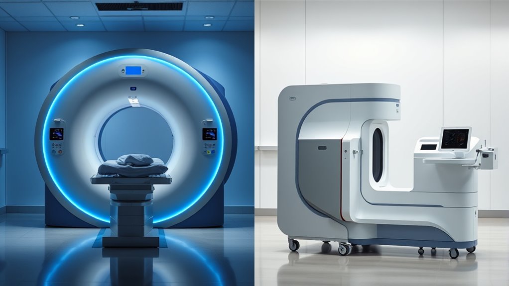
Understanding the costs and equipment needs of CT and cone beam CT is essential when choosing the right imaging option. You’ll want to evaluate factors like initial investment, maintenance, and how accessible each technology is in your setting. Let’s explore how these aspects impact clinical practice and decision-making.
Cost Comparison Factors
Cost is a fundamental factor when choosing between traditional CT and Cone Beam CT for temporal bone imaging, as it directly impacts both healthcare providers and patients. You should consider several aspects:
- Equipment costs: Cone Beam CT machines generally have lower purchase prices compared to traditional CT systems.
- Maintenance expenses: Cone Beam CT units often incur reduced maintenance costs, saving money over time.
- Operational costs: Since Cone Beam CT scans are quicker and often require less staff time, they can lower overall procedure expenses.
- Additionally, the safety profile and radiation exposure levels of Cone Beam CT can influence its suitability, especially in pediatric or sensitive cases, aligning with safety standards and best practices.
While traditional CTs might have higher upfront and ongoing costs, their broader capabilities can justify the investment in certain settings. Balancing these cost factors helps you select the most economical and efficient imaging option.
Equipment Accessibility and Availability
Access to imaging equipment substantially influences the choice between traditional CT and Cone Beam CT for temporal bone imaging. Equipment availability impacts your ability to schedule scans promptly and efficiently. Cone Beam CT units are often more accessible in outpatient clinics due to their smaller size and lower cost, improving facility accessibility. Traditional CT scanners, however, may be limited to larger hospitals or specialized centers, reducing immediate availability. If your facility lacks dedicated imaging equipment, patient access to timely scans may be delayed. Consider the equipment requirements, including space, technical expertise, and maintenance needs, as they directly affect your workflow. Additionally, understanding fetal development and the importance of timely imaging can help prioritize resource allocation. Ultimately, the availability of suitable equipment determines how smoothly you can incorporate either modality into your diagnostic process.
Specific Clinical Scenarios and Use Cases
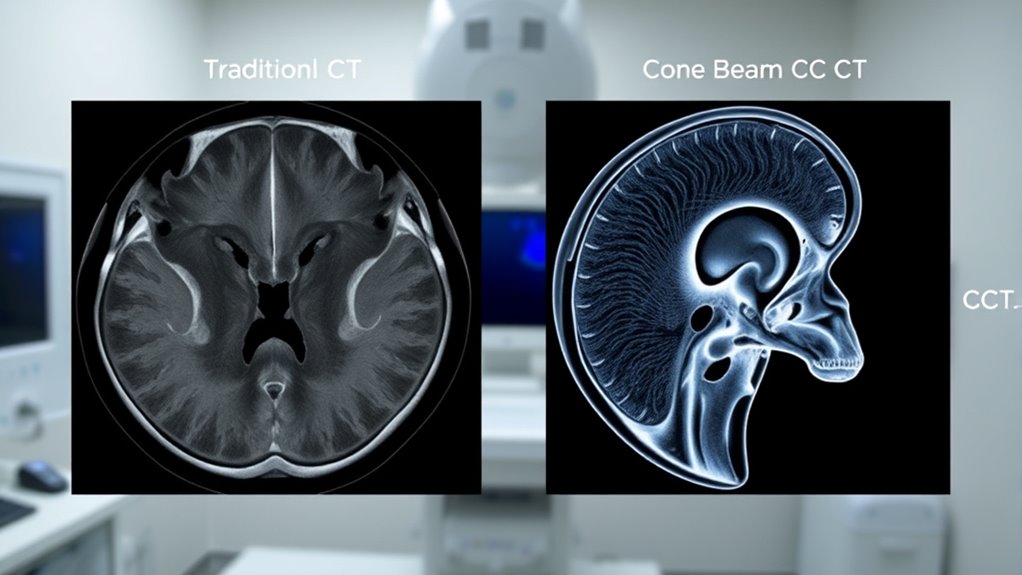
When choosing between CT and Cone Beam CT for temporal bone imaging, clinicians must consider specific clinical scenarios to determine the most suitable modality. For example, in surgical planning, detailed bone structure visualization is essential, making conventional CT preferable. For pediatric imaging, Cone Beam CT offers lower radiation exposure, which is safer for children. Additionally, these scenarios include:
- Preoperative surgical planning for complex ear surgeries where detailed anatomy guides your approach.
- Pediatric cases where minimizing radiation dose is vital without compromising image quality.
- Assessment of congenital anomalies to identify structural deformities early and plan treatment effectively.
Understanding these use cases helps you select the best imaging modality, ensuring accurate diagnosis and patient safety.
Future Trends and Technological Advancements

Advancements in imaging technology are rapidly shaping the future of temporal bone diagnostics, offering new possibilities for more accurate and efficient assessments. AI integration is becoming a game-changer, enabling smarter image analysis, improved detection of subtle abnormalities, and streamlined workflows. This technology allows for real-time interpretation, reducing diagnostic errors and increasing speed. Additionally, miniaturized devices are emerging, making imaging more accessible and patient-friendly. Portable cone beam CT units, for example, can be used in various clinical settings, including outpatient clinics or remote areas. These innovations promise to enhance diagnostic precision, patient comfort, and overall workflow efficiency, ensuring you stay ahead in managing complex temporal bone issues. The future of imaging holds exciting potential for more personalized and precise ear and skull base care. Emerging imaging innovations are also expected to further improve the quality and scope of temporal bone diagnostics.
Frequently Asked Questions
How Do Patient Age and Anatomy Affect Imaging Choice?
When choosing an imaging method, you should consider age-related factors and anatomical variations. Younger patients may have smaller or developing structures that require high-resolution imaging, while older patients might have calcifications or other changes affecting image clarity. Your goal is to select an option that provides detailed visualization of the temporal bone, accounting for these age-related considerations and anatomical variations, ensuring accurate diagnosis and ideal patient care.
Are There Specific Contraindications for CBCT in Temporal Bone Imaging?
When considering CBCT for temporal bone imaging, you should be aware of contraindications like increased radiation exposure risks, especially for pregnant patients or those requiring multiple scans. Allergic reactions are rare but possible if contrast agents are used. Always evaluate the patient’s medical history and specific needs to determine if CBCT is appropriate, ensuring safety and minimizing potential adverse effects.
How Do Different Pathologies Influence Modality Selection?
When selecting an imaging modality, you consider pathology specificity and imaging sensitivity. Different pathologies, like cholesteatoma or ossicular chain issues, require high sensitivity to detect subtle changes. Cone beam CT offers excellent spatial resolution, ideal for detailed bony structures, while conventional CT provides broader tissue contrast. Your choice depends on the pathology’s nature; for soft tissue issues, standard CT might be preferable, whereas for bony details, cone beam CT excels.
What Are the Environmental Impacts of Each Imaging Technique?
Environmental impacts of imaging techniques highlight a stark contrast: while both emit radiation, the radiation footprint of traditional CT is higher, contributing more to environmental concerns. Cone beam CT, with its lower energy consumption, minimizes ecological harm and reduces radiation exposure. You can choose the method that not only offers diagnostic precision but also aligns with sustainable practices, balancing patient safety and environmental responsibility effectively.
Can Portable Imaging Devices Replace Traditional CT or CBCT?
You might wonder if portable imaging devices can replace traditional CT or CBCT. While mobile imaging offers greater convenience and cost-effectiveness, it often lacks the image quality and resolution needed for detailed temporal bone assessments. For accurate diagnosis, especially in complex cases, traditional CT or CBCT remains essential. Portable devices are great for quick screenings but shouldn’t fully replace standard imaging methods when precise, high-quality images are required.
Conclusion
So, whether you prefer the detailed clarity of traditional CT or the convenience of cone beam CT, it seems your choice might just boil down to your patience for radiation exposure or your wallet’s patience for costs. Ironically, the more advanced technology promises better images, yet sometimes, the old-school scan still wins in accessibility. In the end, your clinical needs and safety come first—because nothing says progress like balancing precision with practicality.


