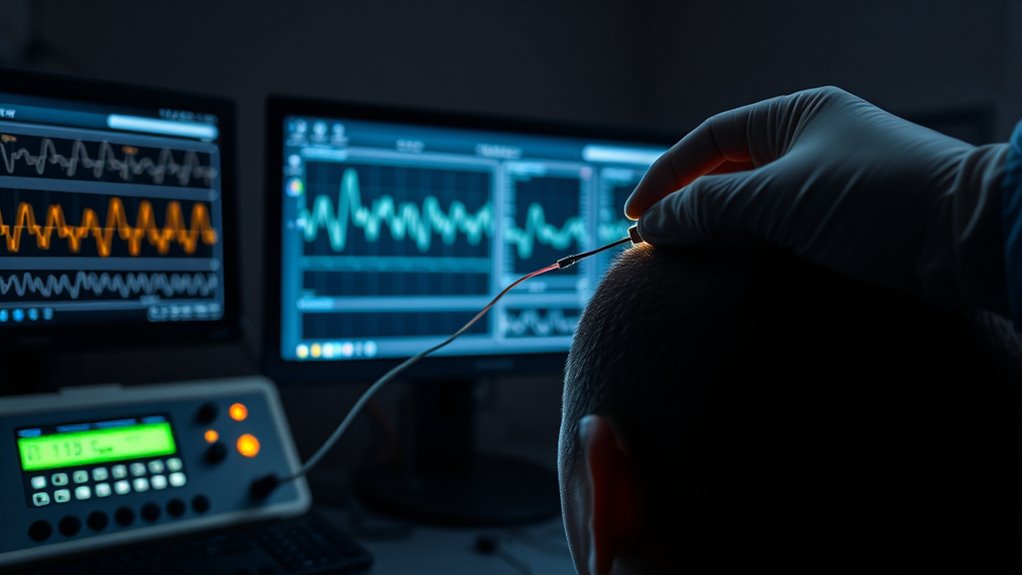
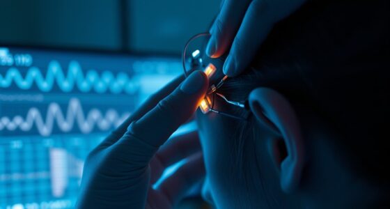
Electrophysiological tests like the Auditory Brainstem Response (ABR) and Envelope Following Response (EFR) help detect hidden hearing loss by measuring neural activity along your auditory pathway. These tests reveal issues like synaptic damage or neural timing problems that standard audiograms might miss, especially when your hearing thresholds appear normal. Keep exploring to learn how these methods uncover subtle auditory nerve issues affecting your hearing quality.
Key Takeaways
- Electrophysiological tests like ABR measure neural responses along the auditory pathway to detect hidden cochlear synaptopathy.
- Envelope Following Response (EFR) assesses neural synchronization, revealing synaptic damage not seen in standard audiograms.
- Middle Latency Responses (MLR) evaluate neural activity 20-80 ms after sound, indicating synaptic integrity and plasticity.
- Advanced techniques utilize high-resolution ABRs and EFRs to identify subtle neural deficits linked to hidden hearing loss.
- These tests provide objective insights into neural communication impairments affecting hearing in noisy environments.
Understanding the Limitations of Standard Hearing Assessments

While standard hearing assessments like audiograms are essential, they often miss subtle forms of hearing loss, such as hidden hearing loss. Noise-induced damage can cause cochlear synaptopathy, where the connections between hair cells and auditory nerve fibers deteriorate. This damage doesn’t usually affect the threshold detected in traditional tests but impairs your ability to understand speech in noisy environments. You might have normal audiogram results yet still struggle to follow conversations in busy settings. Standard tests focus on detecting loudness issues, but cochlear synaptopathy affects neural communication without changing volume thresholds. As a result, many cases of hidden hearing loss go unnoticed without specialized testing, making it vital to understand their limitations in identifying auditory nerve damage caused by noise exposure. Self Watering Plant Pots can serve as an analogy for understanding how some damage occurs internally without immediate visible signs.
Auditory Brainstem Response (ABR) Testing

Auditory Brainstem Response (ABR) testing is a valuable tool for detecting neural issues in the auditory pathway that traditional hearing tests often miss. It measures electrical activity along the auditory nerve and brainstem in response to sound stimuli. When the cochlear synapse or auditory nerve isn’t functioning properly, the ABR can reveal delays or abnormalities in neural signal transmission. During the test, electrodes are placed on your scalp to record responses to brief sounds, such as clicks or tones. If the ABR shows atypical wave patterns or delayed responses, it suggests hidden damage at the cochlear synapse or along the neural pathway. This makes ABR testing especially useful for uncovering neural deficits linked to hidden hearing loss that standard audiograms might overlook. Additionally, understanding neural signal transmission can help interpret the results more accurately and guide appropriate intervention strategies.
Envelope Following Response (EFR) and Its Significance

The Envelope Following Response (EFR) is a specialized electrophysiological test that measures how well your auditory system can track the amplitude changes of complex sounds over time. It reflects neural synchronization in the auditory pathway, which is vital for processing speech and music. EFR is particularly useful for detecting cochlear synaptopathy, a form of hidden hearing loss where synapses between hair cells and auditory nerve fibers are damaged. When these synapses are compromised, your EFR responses weaken, indicating impaired neural synchronization. This test helps reveal hidden deficits not apparent in standard audiograms. Understanding EFR’s significance allows clinicians to identify subtle neural issues affecting your hearing, especially in noisy environments. Here’s a quick overview:
| Aspect | Significance |
|---|---|
| Neural synchronization | Key for processing complex sounds |
| Cochlear synaptopathy | Damage affects EFR responses |
| Diagnostic value | Detects hidden hearing deficits |
Using Middle Latency Responses to Detect Synaptic Damage

Middle Latency Responses (MLRs) offer a valuable window into neural activity occurring roughly 20 to 80 milliseconds after sound presentation, making them especially useful for detecting synaptic damage in the auditory pathway. Damage to synapses can impair synaptic plasticity, reducing the brain’s ability to adapt or reorganize in response to auditory stimuli. MLRs are sensitive to these changes because they reflect neural synchronization within thalamo-cortical circuits. When synaptic connections weaken or become less efficient, MLR amplitudes diminish, indicating disrupted neural timing and coordination. By analyzing these responses, you can identify subtle synaptic damage that traditional audiometry might miss. This approach helps reveal hidden hearing deficits rooted in synaptic dysfunction, providing a more holistic understanding of auditory health. Additionally, understanding the relationship between synaptic integrity and phenomena like hidden hearing loss can aid in developing targeted diagnostic and therapeutic strategies.
Emerging Electrophysiological Techniques in Hidden Hearing Loss Detection

Recent advances in electrophysiological research have introduced innovative techniques that enhance our ability to detect hidden hearing loss beyond traditional methods. These emerging tools leverage our understanding of neural plasticity, allowing us to observe how the auditory system adapts to cochlear synaptopathy. Techniques like expanded auditory brainstem responses (ABRs) and envelope following responses (EFRs) now target subtle neural changes associated with synaptic damage that aren’t visible in standard audiograms. By focusing on neural plasticity, these methods can reveal deficits in synaptic connections between hair cells and auditory nerve fibers. This allows for more precise identification of cochlear synaptopathy, a core component of hidden hearing loss, even when hearing thresholds appear normal. The development of high refresh rates in electrophysiological testing equipment further improves the temporal resolution necessary to detect rapid neural responses associated with early synaptic damage. These advancements promise to improve early detection and intervention strategies in auditory health.
Frequently Asked Questions
Can Electrophysiological Tests Differentiate Between Cochlear and Neural Synaptopathy?
You wonder if electrophysiological tests can differentiate between cochlear and neural synaptopathy. These tests help identify the site of damage by measuring neural responses, which can suggest whether issues stem from cochlear hair cells or synaptic connections. While they provide valuable clues, complete differentiation remains challenging. Still, with careful interpretation, electrophysiological assessments can aid in synaptopathy differentiation, improving diagnosis and targeting treatment strategies effectively.
How Do Age-Related Changes Affect Electrophysiological Responses in Hidden Hearing Loss?
Ever wondered how age-related decline impacts your hearing? As you age, neural degeneration and structural changes can alter electrophysiological responses, making it harder to detect hidden hearing loss. These changes may reduce the amplitude and delay response times, complicating diagnosis. Recognizing these age-related alterations helps you understand that normal test results might not rule out early neural damage, emphasizing the importance of tailored assessment strategies for older adults.
Are There Specific Electrophysiological Markers for Tinnitus Associated With Hidden Hearing Loss?
You’re curious if there are electrophysiological markers for tinnitus linked to hidden hearing loss. Researchers aim for biomarker identification to improve diagnostic specificity, making it easier to distinguish tinnitus caused by hidden hearing loss. Some studies suggest altered auditory brainstem responses or abnormal wave patterns could serve as potential markers. While promising, these markers still need validation for consistent diagnostic use, so ongoing research is essential.
What Are the Limitations of Electrophysiological Testing in Real-World Noisy Environments?
You’ll find that testing limitations in real-world noise can affect your ability to accurately assess hearing. In noisy environments, background sounds interfere with electrophysiological measurements, making it harder to isolate specific neural responses. This means tests may not reflect your everyday hearing challenges accurately. As a result, results can be less reliable, and clinicians may struggle to evaluate your hearing abilities fully outside controlled settings.
How Soon Can Electrophysiological Techniques Detect Early Stages of Synaptic Damage?
You wonder how soon electrophysiological techniques can detect early synaptic damage. These methods offer promising early detection capabilities, often identifying issues before noticeable hearing loss occurs. The diagnostic timing varies but can be quite rapid, sometimes within days or weeks of initial damage. This quick response enables proactive intervention, helping preserve hearing health. However, accuracy depends on the specific technique and the extent of damage, so timely testing is essential.
Conclusion
While standard hearing tests can miss hidden damage, electrophysiological measures like ABR and EFR act as detectives uncovering silent synaptic injuries. These tools shine a light on the unseen, much like a flashlight revealing secrets in the dark. As research advances, you’ll gain sharper insights into hearing health, ensuring no hidden issue slips through unnoticed. Embracing these techniques is like adding a new set of eyes, making sure you catch every whisper of hearing loss.

